New tool navigates the brain’s addiction mechanisms

Opioid addiction is a widespread and complex issue, both in society and in the brain.
Barbara Smith, an assistant professor of biomedical engineering in the Ira A. Fulton Schools of Engineering at Arizona State University, is helping the research community better understand how addiction affects the brain at the cellular level to better combat this debilitating condition.
With support from a 2020 National Science Foundation Faculty Early Career Development Program (CAREER) Award, Smith and her research team are pioneering a new tool to identify and target specific brain cells in a way that has not been possible until now.
“My laboratory is focused on establishing an accurate and scalable technology to better understand fundamental ways the brain communicates across widespread neurocircuitry,” Smith says. “These tools are being developed to explore the underlying mechanisms of opioid addiction and other neurological disorders and diseases.”
 Part of what makes the brain amazing is its neuroplasticity, or its ability to change over the course of our lives. These changes can be positive, such as when abilities are remapped after an area of the brain is damaged by a debilitating event like a stroke.
But neuroplasticity can also be negative. In the case of addiction, the mesolimbic dopamine system — the brain’s reward system — can be reprogrammed in a way that keeps a person dependent on drugs such as opioids.
A certain type of neuron (brain cell) in the mesolimbic dopamine system plays a key role in addiction and programming the brain to view opioid use as rewarding. To find ways to address this, it is necessary to understand the function of those cells, how addiction makes changes in them and how the cells are communicating with other cells in a circuit around different areas of the brain.
“In the CAREER Award project, I propose a new neuronal navigation tool that will enable us to target, modulate and record the activity of interconnected neurons across the brain,” Smith says.
Cells that play a role in addiction are widely spread across different regions of the brain and at different depths. Researchers who want to better understand this illness are limited in their ability to target specific cells of interest. Cells within a couple of millimeters from the surface of the brain can be targeted with current methods. However, researchers have had no way to target specific cell types at depths greater than 2 millimeters if they want high-resolution recordings of the cellular activity. Without these high-resolution recordings, it is difficult to understand what is happening to the cells under the effects of addiction.
Smith and her team are pioneering the development of a first-of-its-kind targeting tool called FLuoro-Acoustic Multipipette Electrodes, or FLAME. The tool improves upon the limitations of existing techniques by incorporating light with acoustics to navigate to specific cell types and get a high-resolution view of what’s happening in those cells, even when they are in deeper regions of the brain.
FLAME integrates a new targeting mechanism into existing micropipette electrodes — tiny glass tubes that have a tip the fraction of the size of a cell. The technology integrates photoacoustics (sound generated by light) and fluorescence (indicators that contrast targets from their surroundings) to navigate toward a specific cell of interest.
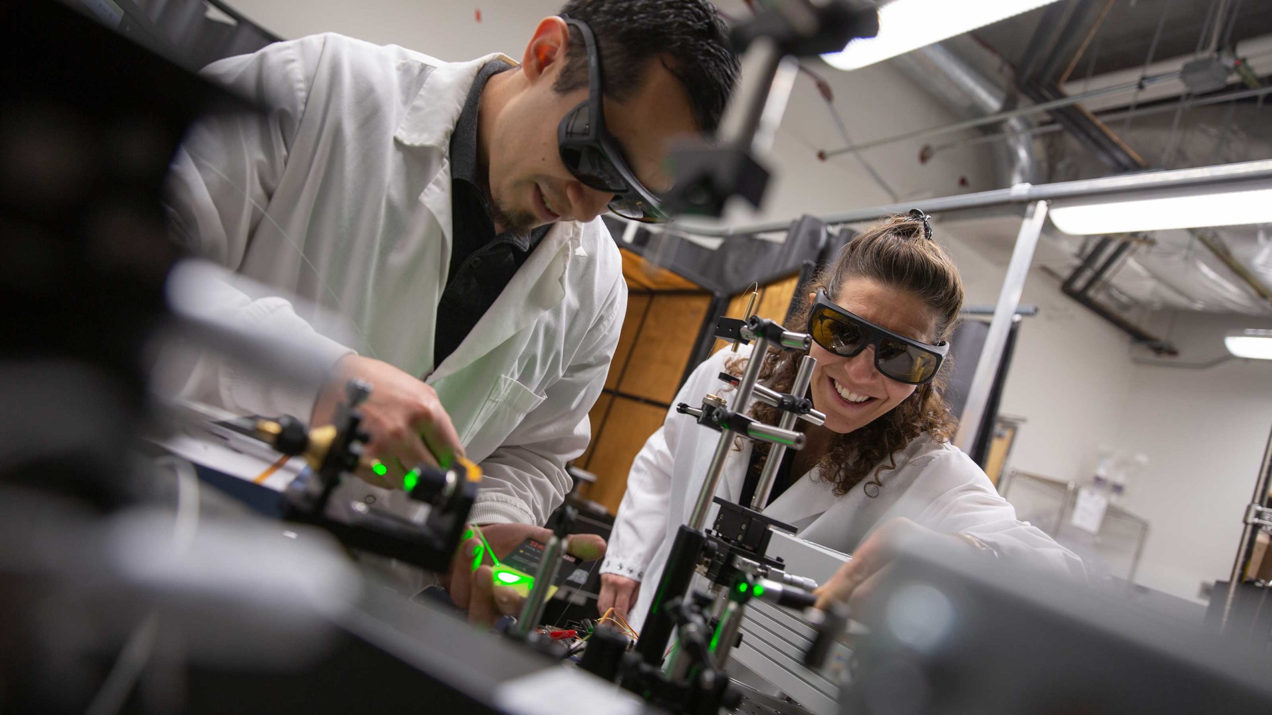
A light, such as a laser, is shone through the micropipette into the brain. The light is absorbed by particular molecules inside of the cell of interest that are genetically marked by photoacoustic and fluorescence indicators. In the case of photoacoustics, the absorbed light results in a pressure wave that is transmitted back up the micropipette and detected as a sound wave. This pressure wave, in combination with fluorescence, relays where the cell is located. Once the cell of interest is identified, FLAME can record its electrical activity at a high resolution.
By efficiently targeting cells of interest and recording their activity, FLAME will help researchers to better understand how addiction affects the brain at a cellular level. This enhanced understanding is an important step to developing preventative medicine and effective treatments for opioid addiction in the future.
Smith’s focus is on interdisciplinary integrated research and education at the intersection of biomedical engineering, neuroscience, biology and medical diagnostics. Her students are developing expertise in molecular biology and imaging methods to expand this research to new depths.
Christopher Miranda, a biomedical engineering doctoral student, is pioneering the development of underlying technology of the FLAME tool. Joel Lusk, a biological design doctoral student, is using molecular biology to develop contrast agents that enable FLAME to target specific cells.
While Smith’s research team is focusing on developing FLAME to study the cellular mechanisms underlying addiction, she says its use could be expanded to study a wide range of neurological diseases and disorders and how they affect the brain’s function at a cellular level.
“Knowledge gained from FLAME will support scientists to make future discoveries within currently unknown areas of neuroscience,” Smith says. “It can open the door to an understanding of the actual workings of deep-brain neurons as they impact addiction, traumatic brain injury, Alzheimer’s disease and pain.”
Monique Clement
Communications specialist, Ira A. Fulton Schools of Engineering
(480) 727-1958 | [email protected]
More news from ASU Engineering

13 ASU engineering faculty earn the prestigious NSF CAREER Award

ASU, Banner Health team up to ease COVID-19 patient isolation

Engineering students supply PPE to health care providers

Shedding new light on nanolasers using 2D semiconductors

Technology engineered at ASU 50 years ago helps battle COVID-19 infection