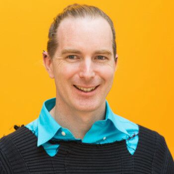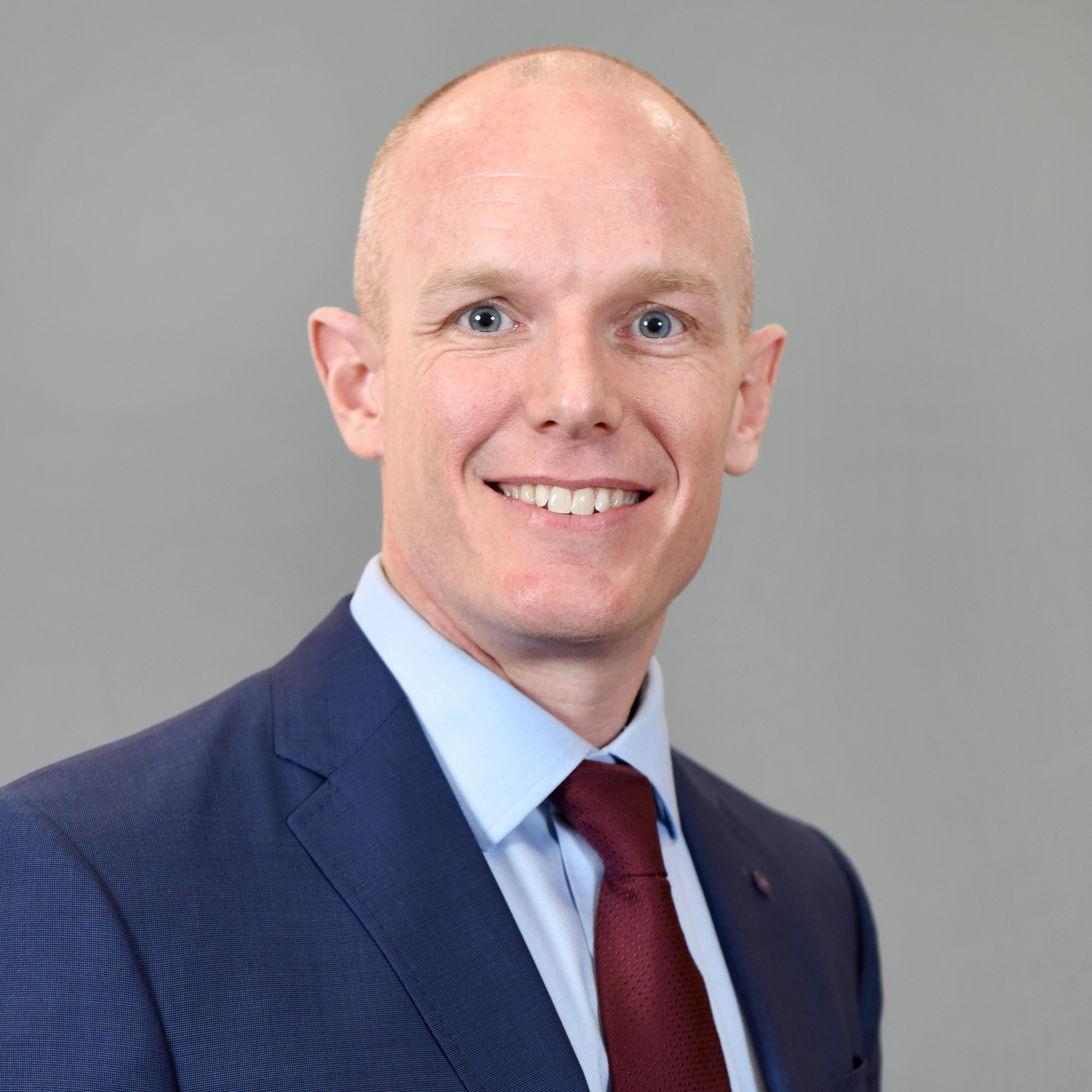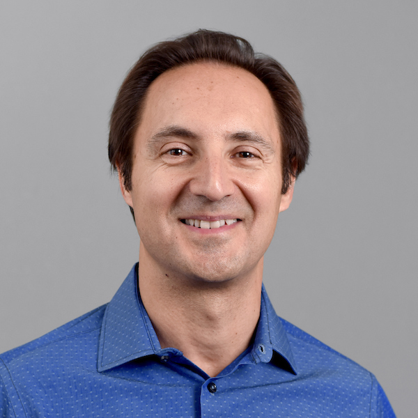

Podcast guests


Podcast host: Lanelle Strawder, Content and Public Relations Manager, Ira A. Fulton Schools of Engineering
Transcript
SPEAKERS
Lanelle Strawder, Scott Beeman, Marco Santello, Craig Smith, Benjamin Bartelle
Lanelle Strawder 00:17
Hello, and welcome to Convergence, the podcast, your audio destination for news and updates about the Ira A. Fulton Schools of Engineering, the largest and most comprehensive engineering school in the United States, where we’re delivering a world class learning experience for students and advancing research and innovation all at scale. I’m your host, Lanelle Strawder, the content and public relations manager for the Fulton Schools of Engineering. Today, we’re back to hear about exciting research news from faculty members in the School of Biological and Health Systems Engineering, one of the seven schools in the Fulton Schools of Engineering.
Taking us on this journey of biomedical engineering innovations is Dr. Marco Santello, a professor and director of the Biological and Health Systems school. Marco joined the University in 1999 and has served in a number of leadership and faculty roles across the university. He’s an expert in soft robotic haptic interfaces that improve sensory motor function. Thank you for joining today, Marco. Can you tell us a little bit more about your school and introduce us to today’s guests?
Marco Santello 1:27
It’s a pleasure to be on this podcast to tell you a little bit about our school, in particular: our faculty. So our school consists of roughly 800 students, roughly 600-650 undergrads and the rest graduate students, split among master and PhD students. We have a number of research thrusts, including neural engineering, synthetic biology, bioimaging and in fact, bioimaging is the area that we’re gonna talk a little bit about today. I have invited two of our assistant professor faculty. One is Dr. Benjamin Bartelle and the second faculty is Dr. Scott Beeman. They are working in complementary areas, although they are both related to bioimaging. Dr. Bartelle, in particular, works on molecular imaging, whereas Scott Beeman works on devising non-invasive magnetic resonance-based methods to quantify tissue microstructure and function.
And we will apply you know, both of their research agendas really look both at the fundamental mechanisms as well, of course, clinical applications. And without further ado, I’d like to give the mic to Dr. Bartelle to tell us a little bit about his work.
Benjamin Bartelle 2:50
Thank you, Marco. I guess I should start out with saying that while people view MRI as a clinical imaging modality, which is fair, it’s one of the most powerful ones you can use, I’m actually, purely a research MRI person, so my subject matter has always been rodents or even just cell samples. And the type of things I work on for MRI usually don’t end up in the clinic or they’re not going to be used on a person.
They’re actually getting at– something for me was the most magical part of MRI coming into it and that’s that you can see all the way through any organism. Yeah, if that’s a person, that’s great, but you have an incredible amount of power if you can do that in a model organism, like a mouse or a rat. We do a lot of molecular techniques on these organisms, namely mice, right? We have these maps of brain expression. We’ve charted out all these cells that they have in this massive cell atlas project, but our ability to look into these animals is actually very limited, right? Some of the best microscope technologies can only get down about a millimeter into a living animal and so that’s not a lot if we’re talking about something the size of a mouse or a rat or organisms of that scale. So for me it started with wanting to see into these animals and the only direction, the only technology available to do that, I thought, was MRI at the time. So for me and my background I sort of focused on well, how do we see gene expression? That would be sort of the first thing I wanted to know about looking into animals. And so my early work was in developing what’s called reporter genes for MRI. Most people know what green fluorescent protein is, that’s a reporter gene for microscopy. So I developed a couple that worked for MRI and that did start out pretty good, but I was convinced upon going into my postdoc that what really mattered was seeing dynamic events, things switching on and off.
And given the signal to noise available to you with MRI as opposed to fluorescents, it actually seemed like a better application. And so, the kind of “second chapter” of my technology development went into making MRI sensors. So we looked at things like serotonin, dopamine… I developed a calcium sensor, which calcium imaging and microscopy is the backbone of modern neuroscience, and that’ll get electrophysiologists mad at me for saying that, but a lot of people use that. So we’re developing that sort of thing for whole brain imaging in animals.
Upon coming here though, it was really time for me to break out on my own and I realized that one thing that we really can’t see by any other means, really, is the molecular signaling of neuro inflammation in neuro immune responses in the brain, right? Lots of people are looking at neurotransmitters and there’s lots of ways of getting at that through other means, but looking at neuro inflammation, for me, that seemed like the ultimate prize.
It’s not something you can get at by other means, right? If you try to use a microscope, generally you notice when you’re opening a hole into your brain to look inside. That’s something that your immune system is pretty wise to– but MRI is totally non-invasive. So for me, that’s my current frontier is how do I pick apart neuro immune and neuro inflammatory signaling going on, right? That could mean things like sensors, building individual sensors or individual molecular technologies, and that’s a big part of my lab, which is the protein engineering aspect, the synthetic biology aspect to try and build these tools.
Another large chunk of that is how do you have those things, right? How do you actually look at those things with MRI? You got to change the hardware and such.
Marco Santello 6:56
So, that’s great. So how do you see the major obstacles on the path to where you want to get to? Meaning, you see technology barriers, you see, you know, other kinds of barriers that can be just biological barriers that it’s difficult to penetrate, how do you see that– your challenges and advancing your area?
Benjamin Bartelle 7:20
Yeah, I see a lot of challenges. A lot of it, for me is just laying out the groundwork being able to “do stuff.” One question, probably the most fundamental biological question is, what signal is inflammation? Is there something that says okay, that’s definitely this particular kind of inflammation? We think of inflammation as like a swollen knee or something, and you take an aspirin and you know, there’s these COX inhibitors gonna end inflammation or an antihistamine can end an allergic response. That’s probably true for those very particular kinds of inflammation, but there’s a wide range of immune challenges in the body. And so what would you look at that would say, this is inflammation? When I first got here, I was convinced like, okay, nitric oxide, that’s a really common inflammatory molecule. That’s definitely the one I want to go for. And I still am, but the deeper I look into it, the more I realize that this is a constellation of different types of molecular signals. And so we may be able to get some information by a single sensor, but we actually need a more integrated approach. Like, that one sensor with other types of imaging around it. And for me, that is also a major bridge to what most people are actually interested in, which is, how does this get to a person? You can build all these sensors and make transgenic animals and have them tell you stuff, but it’s something we’ve learned in the past, say five years or so. And we’ve known for a while, but the past five years are really demonstrating to us discoveries in a mouse, cures in a mouse, they don’t really translate to a person. So how can this work move on to a person? And first, the diagnostic aspect is kind of an obvious connection. And that’s definitely there. Like if you’re going to try to detect something with a sensor in a mouse to get this really high resolution granular understanding of this molecular pathway… what does that mean in a human? Is there some analog? Is there some biomarker you could then look at, for a person where you just can’t make a transgenic person specifically to look at nitric oxide signaling? So, making that jump, I see that as the sort of secondary challenge. So the number one- what do you look at? Number two- how does that relate to a human being? Or, how does that translate into a human? We know neuro immunities are so different between organisms.
Marco Santello 9:52
Thank you. Maybe this is a good segue into Dr. Scott Beeman’s work, as part of his work is really on human and clinical applications for what path of technology development, but nevertheless, it sounds as if Scott’s work, you know, compliments really nicely for the work that Dr. Bartelle does. So, I’m gonna pass the mic now to Dr. Beeman.
Scott Beeman 10:22
Thanks a lot, Marco. Yeah, indeed, that’s a great transition. You know, Ben and I share common goals. However, our approaches are completely different and thus, I’ve always found our work to be totally complementary to one another. We’re kind of working in parallel towards one goal.
That’s kind of the nature of science right? 1000 Projects launch and maybe 10 of them succeed. So I always like taking a step back and looking at where, you know, Ben, myself, Dr. Kodibagkar and the rest of our imaging folks are going. At any rate, the specific goals of my lab are to quantify biologic structure and function. That’s both on the normal physiology side of things, as well as the pathology side of things. So when I say structure, I may mean something to the effect of how big are the cells in this particular tissue? Where’s the basement membrane? And how does it hold things together? Things of that nature.
When I talk about function, I may mean is this tissue being appropriately perfused by blood to deliver the appropriate nutrients and oxygen for the tissue to function? Or are the cells here taking up their nutrients and metabolizing them in the appropriate way? And you can imagine a convergence of both of those things, and often we end up in that gray area. For example, the vasculature is a structure and the flow of the water through that vasculature is a function. And so I’ve kind of fallen in love with the marriage of all of these things and how it propagates itself into that MRI signal.
My background started, you know, I kind of really kind of fell backwards into this field. Strangely enough, I was an undergraduate at ASU back in 2002, which is now a long time ago. And long story short, I was actually a lifeguard and one of my lifeguarding compatriots had a partner who was doing some functional MRI research at the Barrow Neurological Institute in Phoenix. I had no idea what functional MRI was, and I had no idea what the Barrow Neurological Institute was. However, it paid reasonably well and it was within my major, which was biomedical engineering. So I did that for about five years even after I graduated college. I stuck around and worked as a research assistant doing functional MRI and structural MRI to interrogate kind of the pathogenesis of the aging brain, as well as just really acute functional MRI and structural MRI to deliver to the operating room for surgeons to know where to cut and perhaps where to not cut a brain if they have to remove a tumor. And so, that’s kind of where I got my feet wet and I wanted to go to medical school like every biomedical engineering undergrad wants to do at some point, but I was pushed by one of my radiology friends to a faculty who was hiring graduate students at ASU. So I ended up back at ASU to do my PhD and that’s where I just really fell in love with the biophysics of MRI.
For those listening to this program, who have taken organic chemistry, for example, you would know immediately that magnetic resonance carries very, very rich data. This is the key tool, one of the key tools, of the physical chemist, right? You can understand how a molecule moves through space, you can understand the structure of a molecule. And so, what I just kind of really embraced was applying those principles to living systems. Of course, I’m not the first person. I’m about 30 years behind being the first person. However, it’s just fascinating. Every time I get to dig into that signal and say, “the water is moving at this particular rate in healthy tissue. It’s moving at this particular rate in a tumor.” It’s just exciting to be able to peer directly into the body without opening it up, you know, no need to put a probe in there. Just look straight into the tissue and tell either a scientific audience or a clinician what that tissue is doing and why that’s important.
Benjamin Bartelle 15:36
I think that’s something that you and I both have talked about a while. I think of MRI, and the “I” in it is imaging, but there’s so much more data in there. It’s sort of like you’re taking the pulse of like, the quantum universe and you’re just taking a picture of it like from very far away. There was a lot going on in there that’s a) just not human interpretable. We look at these like, spectra, and it’s a mess that we tend to think of as noise. But everything is telling us there is like real molecular information. You can see what molecules are doing what in the brain. There might not be enough of them to reliably tell you something except for a few metabolites, but we can get at it. And I think that’s kind of the direction we’re both headed, which is really making use of that signal, right? There’s this multi-million dollar machine that can go all the way down to just micron or hundreds-of-microns scale in an organism. They can tell what is happening at the molecular level in a functional way. And we just haven’t even touched on that stuff yet as a scientific community. They just barely scratch that surface. I think that’s the whole wide field that both of us kind of talk about, but it’s like how do you get a toehold on all that stuff?
Scott Beeman 16:58
I 100% agree. Marco, I hope I don’t head-off one of the questions, but one of the questions that you asked Ben earlier was, “What kind of challenges are we faced with?” And as engineers, our minds immediately go to what we think of as “technology.” What would commonly be referred to as “technology,” meaning “hardware” and “software,” things of that nature. Boy, there’s a lot of work to be done on that end. However, a lot of the MRI technology is somewhat mature in that regard. There’s fantastic coils out there. Computers are just stellar. They’ve now exceeded our needs, right? However, MRI as a whole is not mature. And that’s exactly to Ben’s point, which is digging deep down, not imaging things on a voxel-wide scale, right? So a cubic millimeter for example, and just say, well, “that’s as good as we can get.” No– we really want to measure things on the molecular scale, on the nanoscopic scale. How far does this water molecule move in a very short amount of time? And what does that tell us about the size of the cell that’s bound within?
Maybe to expand a bit more on what Ben said: I have been working on this analogy for years and never quite, quite clicked in the in the right way, but I’m a terrible photographer, but I like to take pictures. So I like to go out and hike and take a picture of a beautiful mountain. And the way I view the current state of the art in the clinic is simply taking a picture of a beautiful mountain, but that doesn’t tell you a lot about the activity on that mountain, right? It doesn’t tell you a lot about the function of that mountain. How many squirrels are in the trees, right? How many trees are there? What types of trees are growing on that mountain? This is absolutely a possibility with magnetic resonance. It just takes a deep understanding of, in this case, biology, and the MRI signal and how you smash those two things together. And so that’s what I love about it because it’s a never ending battle, right? There’s small wins along the way that makes you feel good and there’s always one step further to go.
Marco Santello 19:45
Well, both of you have done a great job illustrating both the challenges, but also the excitement in making these discoveries. Though, then again as you said, there are still many steps, and in particular, many steps that will eventually come with some clinical application. Nevertheless, it sounds as if you’re developing cutting edge tools with the, you know, high risk, but also high reward kind of questions. So that’s really exciting and I’m very happy that you’re part of the school and also it is definitely complementary in nature as you’ve already explained. You might be using different tools, but at the end of the day you’re going to be converging to common kinds of problems and coming up with solutions.
So let me just end on the on the clinical questions, meaning that you know, some of the listeners might know, even though we have a highly ranked undergraduate and graduate biomedical engineering program, we’re one of very few schools in the country that do not have a medical school on campus. And yet, we’ve been very successful in building very strong relations. So, Scott already mentioned his early exposure, which continues to this day with the Barrow Neurological Institute, which is a world class institute in Phoenix.
The Mayo Clinic was mentioned earlier and I just wanted to point out that we still have bridges and very, very good collaborators in the medical community around us. There are tools to bring the forefront, peoples’ problems, and address, you know, needs of patients that kind of keep us, all of us, “real,” in the sense of reminding us what we do at the end of the day, is to put the human condition first. Even though we’re very interested in basic mechanism, nevertheless, still in the back of our mind, you know, the ultimate goal of raising awareness and improving health in the community.
Lanelle Strawder 21:56
All right, well, thank you. I would like to thank each of our guests for catching us up with your latest research and also telling us a little bit more about the paths forward in your field. So we’re just about out of time, but before we go, I would like to ask each of you: Is there anything else that we haven’t covered that you would like our audience to know?
Benjamin Bartelle 22:18
I guess, if we have students and such listening, I say the big issue I have is people get very intimidated by it. I know that Scott and I’ve been talking about sort of these vague, like, “mysteries of the universe stuff.” And there are ways of accessing it at every level. Like yes, being really great at math definitely helps a lot. I wasn’t good at math ‘til I started using MATLAB and programming languages. I just couldn’t even do three lines of algebra without screwing it up. But once I broke through and got to that level, got to a level where, you know, I found a way of accessing it, it suddenly got really rewarding. So I get that a lot from students where it’s like, “well, I can’t do that” or like, “that’s just too complicated for me to go after.”
I’m here to say it’s actually not. You can use this if you get past that membrane, past that intimidation factor. It’s amazing what you can tap into and the things that you can see that no one else has seen before. It’s just like, just this slight bit of pain to get there, but you can get there.
Scott Beeman 23:27
Yeah, I mean, I guess also speaking to our students if any of you are listening, I like to say this in all my classes that my most prized skill that I’ve developed over the last 20 years is my ability to fail. I’m really good at failing and I really mean that when I say “good,” right? That’s it’s very hard and I think over the years, the vast majority of my career has been spent failing, right? That’s the nature of science and I’m proud of the thick skin that I’ve grown and how I’ve been taught by my mentors to take failures and turn them into future successes. And so I hope to project that upon my graduate students and my undergraduates and in class. I guess since I have the ear of whoever’s listening here, I hope to project that on to you as well.
Marco Santello 24:28
That’s a really nice way of wrapping up this podcast. Perseverance, at the end of the day, the drive to solve problems, and if that’s stronger than the occasional frustration that you may have along the way, then that’s the key I think to make it not just to grad school, but in general. Pretty much any professional in health science. So thank you very much for sharing both your technical expertise, but also the words of wisdom for our students. We appreciate it. Thank you.
Lanelle Strawder 25:03
This has been another excellent foray into the world of biomedical engineering. And thank you for all that great information for students and others who may be interested in entering into the field. We’ll be sure to include contact information for Director Dr. Santello, Dr. Bartelle and Dr. Beeman in the show notes. If you’re interested in learning more about advances in Biological and Health Systems Engineering at ASU, or the work of any of our amazing researchers, please visit us on the web at SBHSE.engineering.asu.edu.
Thank you to our guests, and to you, our listeners for joining us for another episode of Convergence, the podcast. Until next time, I’m Lanelle Strawder signing off from ASU Engineering.